


We make two visits each to Back Pond, Hancock Pond, Highland Lake, Keoka Lake, Keyes Pond, Long Lake (north basin), McWain Pond, Middle Pond, Moose Pond (main basin), Peabody Pond, Sand Pond, Trickey Pond, and Woods Pond. For each lake visit, we traveled by foot over the ice to the deep site and used an ice auger to drill a hole.
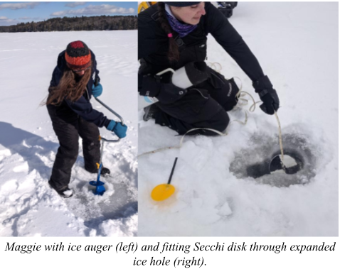
Holes were widened using an ice saw or by drilling an extra hole in order to accommodate larger gear. We used a homemade gauge to measure ice thickness, snow depth, and water level in the hole. We also captured video footage of the ice and under-ice conditions for each lake using a GoPro camera in a waterproof housing. Staff involved in these trips included Maggie Welch, Ben Peierls, Rachel Harper, and Michael Flannery.

We measured light levels above and at several depths below the ice using a LI-COR LI-192 underwater quantum sensor. During these measurements, we covered the hole with three layers of window screening to keep sunlight from passing straight through and affecting the under-ice readings. The attenuation of light due to ice was calculated as the percent of surface light that reaches the water just under the ice layer. Water clarity below ice was measured during each visit using a Secchi disk lowered through the hole and viewed with our standard slanted-glass viewing scope.
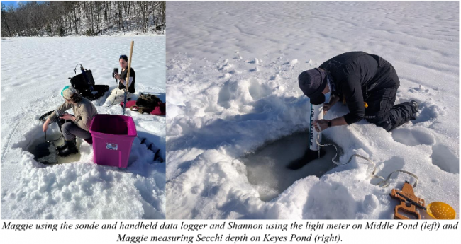
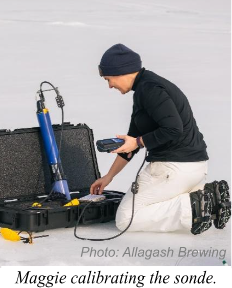
We used a calibrated YSI EXO2 multiparameter sonde connected to a handheld data logger to measure depth profiles of temperature, dissolved oxygen, and chlorophyll fluorescence; the sonde also measures conductivity, pH, and turbidity, but these data are not included in this report. Sonde depth was converted to and reported as depth below ice. Measurements were recorded every 0.5 or 1 meter to the bottom (determined by feel or when turbidity levels rose an order of magnitude).
Water samples were collected using flexible tubing (known as a core tube), which integrates water from the ice to 10 m depth (or to 1 m above the bottom in shallow lakes). These samples were analyzed for total phosphorus using a SEAL segmented flow analyzer, for chlorophyll-a by chemical extraction and fluorescence, and for algae using a Yokogawa Fluid Imaging Technologies FlowCam, a flow imaging microscope that captures images of algae for counting and identification.

